Oncology
Three-dimensional imaging of upper tract urothelial carcinoma improves diagnostic yield and accuracy
Abstract
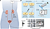
Upper tract urothelial carcinoma (UTUC) is a rare form of urothelial cancer with a high incidence of recurrence and a low survival rate. Almost two-thirds of UTUCs are invasive at the time of diagnosis; therefore, improving diagnostic methods is key to increasing survival rates. Histopathological analysis of UTUC is essential for diagnosis and typically requires endoscopy biopsy, tissue sectioning, and labeling. However, endoscopy biopsies are minute, and it is challenging to cut into thin sections for conventional histopathology; this complicates diagnosis. Here, we used volumetric 3-dimensional (3D) imaging to explore the inner landscape of clinical UTUC biopsies, without sectioning, revealing that 3D analysis of phosphorylated ribosomal protein S6 (pS6) could predict tumor grade and prognosis with improved accuracy. By visualizing the tumor vasculature, we discovered that pS6+ cells were localized near blood vessels at significantly higher levels in high-grade tumors than in low-grade tumors. Furthermore, the clustering of pS6+ cells was associated with shorter relapse-free survival. Our results demonstrate that 3D volume imaging of the structural niches of pS6 cells deep inside the UTUC samples improved diagnostic yield, grading, and prognosis prediction.
Authors
Keishiro Fukumoto, Kanatani, Jaremko, West, Li, Takamatsu, Al Rayyes, Mikami, Niwa, Andri Axelsson, Tanaka, Oya, Miyakawa, Brehmer, Per Uhlén

Abstract
Radiation therapy (RT) is frequently used to treat cancers, including soft-tissue sarcomas. Prior studies established that the toll-like receptor 9 (TLR9) agonist cytosine-phosphate-guanine oligodeoxynucleotide (CpG) enhances the response to RT in transplanted tumors, but the mechanisms of this enhancement remain unclear. Here, we used CRISPR/Cas9 and the chemical carcinogen 3-methylcholanthrene (MCA) to generate autochthonous soft-tissue sarcomas with high tumor mutation burden. Treatment with a single fraction of 20 Gy RT and 2 doses of CpG significantly enhanced tumor response, which was abrogated by genetic or immunodepletion of CD8+ T cells. To characterize the immune response to CpG+RT, we performed bulk RNA-Seq, single-cell RNA-Seq, and mass cytometry. Sarcomas treated with 20 Gy and CpG demonstrated increased CD8 T cells expressing markers associated with activation and proliferation, such as Granzyme B, Ki-67, and IFN-γ. CpG+RT also upregulated antigen presentation pathways on myeloid cells. Furthermore, in sarcomas treated with CpG+RT, TCR clonality analysis suggests an increase in clonal T cell dominance. Collectively, these findings demonstrate that CpG+RT significantly delays tumor growth in a CD8 T cell–dependent manner. These results provide a strong rationale for clinical trials evaluating CpG or other TLR9 agonists with RT in patients with soft-tissue sarcoma.
Authors
Chang Su, Collin L. Kent, Matthew Pierpoint, Warren Floyd, Lixia Luo, Nerissa T. Williams, Yan Ma, Brian Peng, Alexander L. Lazarides, Ajay Subramanian, Jonathon E. Himes, Vincent M. Perez, Rosa D. Hernansaiz-Ballesteros, Kimberly E. Roche, Jennifer L. Modliszewski, Sara R. Selitsky, Mari L. Shinohara, Amy J. Wisdom, Everett J. Moding, Yvonne M. Mowery, David G. Kirsch
Abstract
Carcinomas are common in humans but rare among closely related “great apes”. Plausible explanations, including human-specific genomic alterations affecting the biology of sialic acids are proposed, but causality remains unproven. Here, an integrated evolutionary genetics-phenome-transcriptome approach studied the role of SIGLEC12 gene (encodes Siglec-XII) on epithelial transformation and cancer. Exogenous expression of the protein in cell lines and genetically engineered mice recapitulated ~30% of the human population in whom the protein is expressed in a form that cannot bind ligand due to a fixed, homozygous, human-universal missense mutation. Siglec-XII null cells/mice recapitulated the remaining ~70% of the human population in whom an additional polymorphic frameshift mutation eliminates the entire protein. Siglec-XII expression drove several pro-oncogenic phenotypes in cell lines, and increased tumor burden in mice challenged with chemical carcinogen and inflammation. Transcriptomic studies yielded a 29-gene signature of Siglec-XII-positive disease and when used as a computational tool for navigating human datasets, pinpointed with surprising precision that SIGLEC12 expression (model) recapitulates a very specific type of colorectal carcinomas (disease) that is associated with mismatch-repair defects and inflammation, disproportionately affects European-Americans, and carries a better prognosis. They revealed a hitherto unknown evolutionary genetic mechanism for an ethnic/environmental predisposition of carcinogenesis.
Authors
Hector A. Cuello, Saptarshi Sinha, Andrea L. Verhagen, Nissi Varki, Ajit Varki, Pradipta Ghosh
Abstract
The regulated glycosylation of the proteome has widespread effects on biological processes that cancer cells can exploit. Expression of N-acetylglucosaminyltransferase V (encoded by Mgat5 or GnT-V), which catalyzes the addition of β1,6-linked N-acetylglucosamine to form complex N-glycans, has been linked to tumor growth and metastasis across tumor types. Using a panel of murine pancreatic ductal adenocarcinoma (PDAC) clonal cell lines that recapitulate the immune heterogeneity of PDAC, we found that Mgat5 is required for tumor growth in vivo but not in vitro. Loss of Mgat5 results in tumor clearance that is dependent on T cells and dendritic cells, with NK cells playing an early role. Analysis of extrinsic cell death pathways revealed Mgat5-deficient cells have increased sensitivity to cell death mediated by the TNF superfamily, a property that was shared with other non-PDAC Mgat5-deficient cell lines. Finally, Mgat5 knockout in an immunotherapy-resistant PDAC line significantly decreased tumor growth and increased survival upon immune checkpoint blockade. These findings demonstrate a role for N-glycosylation in regulating the sensitivity of cancer cells to T cell killing through classical cell death pathways.
Authors
Erin E. Hollander, Rosemary E. Flock, Jayne C. McDevitt, William P. Vostrejs, Sydney L. Campbell, Margo I. Orlen, Samantha B. Kemp, Benjamin M. Kahn, Kathryn E. Wellen, Il-Kyu Kim, Ben Z. Stanger

Abstract
Immune therapy is the new frontier of cancer treatment. Therapeutic radiation is a known inducer of immune response and can be limited by immunosuppressive mediators including cyclooxygenase-2 (COX2) that is highly expressed in aggressive triple negative breast cancer (TNBC). A clinical cohort of TNBC tumors revealed poor radiation therapeutic efficacy in tumors expressing high COX2. Herein, we show that radiation combined with adjuvant NSAID (indomethacin) treatment provides a powerful combination to reduce both primary tumor growth and lung metastasis in aggressive 4T1 TNBC tumors, which occurs in part through increased antitumor immune response. Spatial immunological changes including augmented lymphoid infiltration into the tumor epithelium and locally increased cGAS/STING1 and type I IFN gene expression were observed in radiation-indomethacin–treated 4T1 tumors. Thus, radiation and adjuvant NSAID treatment shifts “immune desert phenotypes” toward antitumor M1/TH1 immune mediators in these immunologically challenging tumors. Importantly, radiation-indomethacin combination treatment improved local control of the primary lesion, reduced metastatic burden, and increased median survival when compared with radiation treatment alone. These results show that clinically available NSAIDs can improve radiation therapeutic efficacy through increased antitumor immune response and augmented local generation of cGAS/STING1 and type I IFNs.
Authors
Lisa A. Ridnour, Robert Y.S. Cheng, Noemi Kedei, Veena Somasundaram, Dibyangana D. Bhattacharyya, Debashree Basudhar, Adelaide L. Wink, Abigail J. Walke, Caleb Kim, William F. Heinz, Elijah F. Edmondson, Donna O. Butcher, Andrew C. Warner, Tiffany H. Dorsey, Milind Pore, Robert J. Kinders, Stanley Lipkowitz, Richard J. Bryant, Jens Rittscher, Stephen T.C. Wong, Stephen M. Hewitt, Jenny C. Chang, Aliaa Shalaby, Grace M. Callagy, Sharon A. Glynn, Stefan Ambs, Stephen K. Anderson, Daniel W. McVicar, Stephen J. Lockett, David A. Wink
Abstract
TTK (MPS1) spindle assembly checkpoint kinase is an emerging cancer target. This preclinical study explored the anti-tumor mechanism of TTK inhibitor OSU13 to define a strategy for clinical development. We observed prominent anti-tumor activity of OSU13 in melanoma, colon, and breast cancer cells, melanoma patient-derived organoids, and mice bearing colon tumors associated with G2 cell cycle arrest, senescence, and apoptosis. OSU13-treated cells displayed DNA damage and micronuclei that triggered the cytosolic DNA-sensing cGAS-STING pathway. STING was required for the induction of several proteins involved in T cell recruitment and activity. Tumors from OSU13-treated mice showed an increased proportion of T and NK cells and evidence of PD-1/PD-L1 immune checkpoint activation. Combining a low-toxicity dose of OSU13 with anti-PD1 checkpoint blockade resulted in prominent STING- and CD8 T cell-dependent tumor inhibition and improved survival. These findings provide a rationale for utilizing TTK inhibitors in combination with immunotherapy in STING-proficient tumors.
Authors
Vijaya Bharti, Amrendra Kumar, Yinchong Wang, Nikhil Roychowdhury, Daniel de Lima Bellan, Beimnet B. Kassaye, Reese Watkins, Marina Capece, Catherine G. Chung, Gerard Hilinski, Anna E. Vilgelm
Abstract
Loss of ferroptosis contributes to the development of human cancer, and restoration of ferroptosis has been demonstrated as a potential therapeutic strategy in cancer treatment. However, the mechanisms of how ferroptosis escape contributes to ovarian cancer (OV) development are not well elucidated. Here we show that ferroptosis negative regulation (FNR) signatures correlated with the tumorigenesis of OV and were associated with poor prognosis, suggesting that restoration of ferroptosis represents a potential therapeutic strategy in OV. High throughput drug screening with a kinase inhibitor library identified MEK inhibitors as ferroptosis inducers in OV cells. We further demonstrated that MEK inhibitor resistant OV cells were less vulnerable to trametinib-induced ferroptosis. Mechanistically, mTOR/4EBP1 signaling promoted SLC7A11 protein synthesis, leading to ferroptosis inhibition in MEK inhibitor resistant cells. Dual inhibition of MEK and mTOR/4EBP1 signaling restrained the protein synthesis of SLC7A11 via suppression of the mTOR-4EBP1 activity to reactivate ferroptosis in resistant cells. Together, these findings provide a promising therapeutic option for OV treatment through ferroptosis restoration by the combined inhibition of MEK and mTOR/4EBP1 pathways.
Authors
Jiaxin Yin, Jianfeng Chen, Jing Han Hong, Yulin Huang, Rong Xiao, Shini Liu, Peng Deng, Yichen Sun, Kelila Xin Ye Chai, Xian Zeng, Jason Yongsheng Chan, Peiyong Guan, Yali Wang, Peili Wang, Chongjie Tong, Qiang Yu, Xiaojun Xia, Choon Kiat Ong, Bin Tean Teh, Ying Xiong, Jing Tan
Abstract
The non-physiological nutrient levels found in traditional culture media have been shown to affect numerous aspects of cancer cell physiology, including how cells respond to certain therapeutic agents. Here, we comprehensively evaluated how physiological nutrient levels impact therapeutic response by performing drug screening in human plasma-like medium (HPLM). We observed dramatic nutrient-dependent changes in sensitivity to a variety of FDA-approved and clinically trialed compounds including rigosertib, an experimental cancer therapeutic that has recently failed in phase 3 clinical trials. Mechanistically, we found that the ability of rigosertib to destabilize microtubules is strongly inhibited by the purine metabolism end product uric acid, which is uniquely abundant in humans relative to traditional in vitro and in vivo cancer models. These results demonstrate the broad and dramatic effects nutrient levels can have on drug response, and how incorporation of human-specific physiological nutrient media might help to identify compounds whose efficacy could be impacted in humans.
Authors
Vipin Rawat, Patrick DeLear, Prarthana Prashanth, Mete Emir Ozgurses, Anteneh Tebeje, Philippa A. Burns, Kelly O. Conger, Christopher Solís, Yasir Hasnain, Anna Novikova, Jennifer E. Endress, Paloma González-Sánchez, Wentao Dong, Greg Stephanopoulos, Gina M. DeNicola, Isaac Harris, David Sept, Frank M. Mason, Jonathan L. Coloff
Abstract
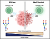
Tumor evolution is driven by genetic variation; however, it is the tumor microenvironment (TME) that provides the selective pressure contributing to evolution in cancer. Despite high histopathological heterogeneity within glioblastoma (GBM), the most aggressive brain tumor, the interactions between the genetically distinct GBM cells and the surrounding TME are not fully understood. To address this, we analyzed matched primary and recurrent GBM archival tumor tissues with imaging-based techniques aimed to simultaneously evaluate tumor tissues for presence of hypoxic, angiogenic, and inflammatory niches, extracellular matrix organization, TERT promoter mutational status, and several oncogenic amplifications on the same slide and location. We found that the relationships between genetic and TME diversity are different in primary and matched recurrent tumors. Interestingly, the texture of the extracellular matrix (ECM), identified by label-free reflectance imaging, was predictive of single-cell genetic traits present in the tissue. Moreover, reflectance of ECM revealed structured organization of the perivascular niche in recurrent GBM, enriched in immunosuppressive macrophages. Single-cell spatial transcriptomics further confirmed the presence of the niche-specific macrophage populations and identified interactions between endothelial cells, perivascular fibroblasts, and immunosuppressive macrophages. Our results underscore the importance of GBM tissue organization in tumor evolution and highlight novel genetic and spatial dependencies.
Authors
Ugoma Onubogu, Chandler D. Gatenbee, Sandhya Prabhakaran, Kelsey Wolfe, Benjamin Oakes, Roberto Salatino, Rachael Vaubel, Oszkar Szentirmai, Alexander R. A. Anderson, Michalina Janiszewska
Abstract
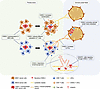
Because cancer cells have a genetically unstable nature, they give rise to genetically different variant subclones inside a single tumor. Understanding cancer heterogeneity and subclone characteristics is crucial for developing more efficacious therapies. Oral squamous cell carcinoma (OSCC) is characterized by high heterogeneity and plasticity. On the other hand, CX3C motif ligand 1 (CX3CL1) is a double-faced chemokine with anti- and pro-tumor functions. Our study reported that CX3CL1 functioned differently in tumors with different cancer phenotypes, both in vivo and in vitro. Mouse OSCC 1 (MOC1) and MOC2 cells responded similarly to CX3CL1 in vitro. However, in vivo, CX3CL1 increased keratinization in indolent MOC1 cancer, while CX3CL1 promoted cervical lymphatic metastasis in aggressive MOC2 cancer. These outcomes were due to double-faced CX3CL1 effects on different immune microenvironments indolent and aggressive cancer created. Furthermore, we established that CX3CL1 promoted cancer metastasis via the lymphatic pathway by stimulating lymphangiogenesis and transendothelial migration of lymph-circulating tumor cells. CX3CL1 enrichment in lymphatic metastasis tissues was observed in aggressive murine and human cell lines. OSCC patient samples with CX3CL1 enrichment exhibited a strong correlation with lower overall survival rates and higher recurrence and distant metastasis rates. In conclusion, CX3CL1 is a pivotal factor that stimulates the metastasis of aggressive cancer subclones within the heterogeneous tumors to metastasize, and our study demonstrates the prognostic value of CX3CL1 enrichment in long-term monitoring in OSCC.
Authors
Htoo Shwe Eain, Hotaka Kawai, Masaaki Nakayama, May Wathone Oo, Toshiaki Ohara, Yoko Fukuhara, Kiyofumi Takabatake, Quisheng Shan, Yamin Soe, Kisho Ono, Keisuke Nakano, Nobuyoshi Mizukawa, Seiji Iida, Hitoshi Nagatsuka
No posts were found with this tag.


