Infectious disease
Abstract
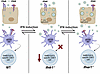
Optimization of protective immune responses against SARS-CoV-2 remains an urgent worldwide priority. In this regard, type III IFN (IFN-λ) restricts SARS-CoV-2 infection in vitro, and treatment with IFN-λ limits infection, inflammation, and pathogenesis in murine models. Furthermore, IFN-λ has been developed for clinical use to limit COVID-19 severity. However, whether endogenous IFN-λ signaling has an effect on SARS-CoV-2 antiviral immunity and long-term immune protection in vivo is unknown. In this study, we identified a requirement for IFN-λ signaling in promoting viral clearance and protective immune programming in SARS-CoV-2 infection of mice. Expression of both IFN and IFN-stimulated gene (ISG) in the lungs were minimally affected by the absence of IFN-λ signaling and correlated with transient increases in viral titers. We found that IFN-λ supported the generation of protective CD8 T cell responses against SARS-CoV-2 by facilitating accumulation of CD103+ DC in lung draining lymph nodes (dLN). IFN-λ signaling specifically in DCs promoted the upregulation of costimulatory molecules and the proliferation of CD8 T cells. Intriguingly, antigen-specific CD8 T cell immunity to SARS-CoV-2 was independent of type I IFN signaling, revealing a nonredundant function of IFN-λ. Overall, these studies demonstrate a critical role for IFN-λ in protective innate and adaptive immunity upon infection with SARS-CoV-2 and suggest that IFN-λ serves as an immune adjuvant to support CD8 T cell immunity.
Authors
Abigail D. Solstad, Parker J. Denz, Adam D. Kenney, Najmus S. Mahfooz, Samuel Speaks, Qiaoke Gong, Richard T. Robinson, Matthew E. Long, Adriana Forero, Jacob S. Yount, Emily A. Hemann
Abstract
Upon infection, naïve CD8+ T cells differentiate into cytotoxic effector cells to eliminate the pathogen-infected cells. Although many mechanisms underlying this process have been demonstrated, the regulatory role of chromatin remodel system in this process remains largely unknown. Here we showed that BRD7, a component of the polybromo-associated BRG1-associated factor complex (PBAF), was required for naïve CD8+ T cells to differentiate into functional short-lived effector cells (SLECs) in response to acute infections caused by influenza virus or lymphocytic choriomeningitis virus (LCMV). BRD7-deficiency in CD8+ T cells resulted in profound defects in effector population and functions, thereby impairing viral clearance and host recovery. Further mechanical studies indicated that the expression of BRD7 significantly turned to high from naïve CD8+ T cells to effector cells, bridged BRG1 and PBRM1 to the core module of PBAF complex, consequently facilitating the assembly of PBAF complex rather than BAF complex in the effector cells. The PBAF complex changed the chromatin accessibility at the loci of Tbx21 gene and up-regulated its expression, leading to the maturation of effector T cells. Our research confirms BRD7 and the PBAF complex are key in CD8+ T cell development and present a significant target for advancing immune therapies.
Authors
Feng Huang, Yingtong Lin, Yidan Qiao, Yaochang Yuan, Zhihan Zhong, Baohong Luo, Yating Wu, Jun Liu, Jingliang Chen, Wanying Zhang, Hui Zhang, Bingfeng Liu
Abstract
Men who have sex with men (MSM) with HIV are at high risk for squamous intraepithelial lesion (SIL) and anal cancer. Identifying local immunological mechanisms involved in the development of anal dysplasia could aid treatment and diagnostics. Here we studied 111 anal biopsies obtained from 101 MSM with HIV, who participated in an anal screening program. We first assessed multiple immune subsets by flow cytometry, in addition to histological examination, in a discovery cohort (n = 54). Selected molecules were further evaluated by immunohistochemistry in a validation cohort (n = 47). Pathological samples were characterized by the presence of Resident Memory T cells with low expression of CD103 and by changes in Natural Killer cell subsets, affecting residency and activation. Furthermore, potentially immune suppressive subsets, including CD15+CD16+ mature neutrophils, gradually increased as the anal lesion progressed. Immunohistochemistry confirmed the association between the presence of CD15 in the epithelium and SIL diagnosis, with a sensitivity of 80% and specificity of 71% (AUC 0.762) for the correlation with high-grade SIL. A complex immunological environment with imbalanced proportions of resident effectors and immune suppressive subsets characterizes pathological samples. Neutrophil infiltration, determined by CD15 staining, may represent a valuable pathological marker associated with the grade of dysplasia.
Authors
Joaquín Burgos, Aleix Benítez-Martínez, Cristina Mancebo-Pérez, Nuria Massana, Antonio Astorga-Gamaza, Josep Castellvi, Stefania Landolfi, Adrià Curran, Jorge N. Garcia-Perez, Vicenç Falcó, María J. Buzón, Meritxell Genescà
Abstract
Since its emergence, SARS-CoV-2 has been continuously evolving, hampering the effectiveness of current vaccines against COVID-19. mAbs can be used to treat patients at risk of severe COVID-19. Thus, the development of broadly protective mAbs and an understanding of the underlying protective mechanisms are of great importance. Here, we isolated mAbs from donors with breakthrough infection with Omicron subvariants using a single–B cell screening platform. We identified a mAb, O5C2, which possesses broad-spectrum neutralization and antibody-dependent cell-mediated cytotoxic activities against SARS-CoV-2 variants, including EG.5.1. Single-particle analysis by cryo-electron microscopy revealed that O5C2 targeted an unusually large epitope within the receptor-binding domain of spike protein that overlapped with the angiotensin-converting enzyme 2 binding interface. Furthermore, O5C2 effectively protected against BA.5 Omicron infection in vivo by mediating changes in transcriptomes enriched in genes involved in apoptosis and interferon responses. Our findings provide insights into the development of pan-protective mAbs against SARS-CoV-2.
Authors
Yi-Hsuan Chang, Min-Feng Hsu, Wei-Nan Chen, Min-Hao Wu, Wye-Lup Kong, Mei-Yeh Jade Lu, Chih-Heng Huang, Fang-Ju Chang, Lan-Yi Chang, Ho-Yang Tsai, Chao-Ping Tung, Jou-Hui Yu, Yali Kuo, Yu-Chi Chou, Li-Yang Bai, Yuan-Chih Chang, An-Yu Chen, Cheng-Cheung Chen, Yi-Hua Chen, Chun-Che Liao, Chih-Shin Chang, Jian-Jong Liang, Yi-Ling Lin, Takashi Angata, Shang-Te Danny Hsu, Kuo-I Lin

Abstract
T cells are required for protective immunity against Mycobacterium tuberculosis. We recently described a cohort of Ugandan household contacts of tuberculosis cases who appear to “resist” M. tuberculosis infection (resisters; RSTRs) and showed that these individuals harbor IFN-γ–independent T cell responses to M. tuberculosis–specific peptide antigens. However, T cells also recognize nonprotein antigens via antigen-presenting systems that are independent of genetic background, known as donor-unrestricted T cells (DURTs). We used tetramer staining and flow cytometry to characterize the association between DURTs and “resistance” to M. tuberculosis infection. Peripheral blood frequencies of most DURT subsets were comparable between RSTRs and latently infected controls (LTBIs). However, we observed a 1.65-fold increase in frequency of MR1-restricted T (MR1T) cells among RSTRs in comparison with LTBIs. Single-cell RNA sequencing of 18,251 MR1T cells sorted from 8 donors revealed 5,150 clonotypes that expressed a common transcriptional program, the majority of which were private. Sequencing of the T cell receptor α/T cell receptor δ (TCRα/δ) repertoire revealed several DURT clonotypes were expanded among RSTRs, including 2 MR1T clonotypes that recognized mycobacteria-infected cells in a TCR-dependent manner. Overall, our data reveal unexpected donor-specific diversity in the TCR repertoire of human MR1T cells as well as associations between mycobacteria-reactive MR1T clonotypes and resistance to M. tuberculosis infection.
Authors
Deborah L. Cross, Erik D. Layton, Krystle K.Q. Yu, Malisa T. Smith, Melissa S. Aguilar, Shamin Li, Elise C. Wilcox, Aude G. Chapuis, Harriet Mayanja-Kizza, Catherine M. Stein, W. Henry Boom, Thomas R. Hawn, Philip Bradley, Evan W. Newell, Chetan Seshadri
Abstract
Inhibition of Bruton's tyrosine kinase (BTK) through covalent modifications of its active site (e.g., ibrutinib [IBT]) is a preferred treatment for multiple B cell malignancies. However, IBT-treated patients are more susceptible to invasive fungal infections, although the mechanism is poorly understood. Neutrophils are the primary line of defense against these infections; therefore, we examined the impact of IBT on primary human neutrophil effector activity against Aspergillus fumigatus. IBT significantly impaired the ability of neutrophils to kill A. fumigatus and potently inhibited reactive oxygen species (ROS) production, chemotaxis, and phagocytosis. Importantly, exogenous TNFα fully compensated for defects imposed by IBT and newer-generation BTK inhibitors and restored the ability of neutrophils to contain A. fumigatus hyphal growth. Blocking TNFα did not impact ROS production in healthy neutrophils but prevented exogenous TNFα from rescuing the phenotype of IBT-treated neutrophils. The restorative capacity of TNFα was independent of transcription. Moreover, the addition of TNFα immediately rescued ROS production in IBT-treated neutrophils indicating that TNFα worked through a BTK-independent signaling pathway. Finally, TNFα restored effector activity of primary neutrophils from patients on IBT therapy. Altogether, our data indicate that TNFα rescues the antifungal immunity block imposed by inhibition of BTK in primary human neutrophils.
Authors
Diego A. Vargas-Blanco, Olivia W. Hepworth, Kyle J. Basham, Patricia Simaku, Arianne J. Crossen, Kyle D. Timmer, Alex Hopke, Hannah Brown Harding, Steven R. Vandal, Kirstine N. Jensen, Daniel J. Floyd, Jennifer L. Reedy, Christopher Reardon, Michael K. Mansour, Rebecca A. Ward, Daniel Irimia, Jeremy S. Abramson, Jatin M. Vyas
Abstract
A systems analysis was conducted to determine the potential molecular mechanisms underlying differential immunogenicity and protective efficacy results of a clinical trial of the radiation-attenuated whole sporozoite PfSPZ Vaccine in African infants. Innate immune activation and myeloid signatures at pre-vaccination baseline correlated with protection from Pf parasitemia in placebo controls. These same signatures were associated with susceptibility to parasitemia among infants who received the highest and most protective PfSPZ Vaccine dose. Machine learning identified spliceosome, proteosome, and resting dendritic cell signatures as pre-vaccination features predictive of protection after highest-dose PfSPZ vaccination, whereas baseline CSP-specific IgG predicted non-protection. Pre-vaccination innate inflammatory and myeloid signatures were associated with higher sporozoite-specific IgG Ab response but undetectable PfSPZ-specific CD8+ T-cell responses post-vaccination. Consistent with these human data, innate stimulation in vivo conferred protection against infection by sporozoite injection in malaria-naïve mice while diminishing the CD8+ T-cell response to radiation-attenuated sporozoites. These data suggest a dichotomous role of innate stimulation for malaria protection and induction of protective immunity of whole-sporozoite malaria vaccines. The uncoupling of vaccine-induced protective immunity achieved by Abs from more protective CD8+ T cell responses suggest that PfSPZ Vaccine efficacy in malaria-endemic settings may be constrained by opposing antigen presentation pathways.
Authors
Leetah Senkpeil, Jyoti Bhardwaj, Morgan R. Little, Prasida Holla, Aditi Upadhye, Elizabeth M. Fusco, Phillip A. Swanson II, Ryan E. Wiegand, Michael D. Macklin, Kevin Bi, Barbara J. Flynn, Ayako Yamamoto, Erik L. Gaskin, D. Noah Sather, Adrian L. Oblak, Edward Simpson, Hongyu Gao, W. Nicholas Haining, Kathleen B. Yates, Xiaowen Liu, Tooba Murshedkar, Thomas L. Richie, B. Kim Lee Sim, Kephas Otieno, Simon Kariuki, Xiaoling Xuei, Yunlong Liu, Rafael B. Polidoro, Stephen L. Hoffman, Martina Oneko, Laura C. Steinhardt, Nathan W. Schmidt, Robert A. Seder, Tuan M. Tran
Abstract
BACKGROUND Persistent cough and dyspnea are prominent features of post-acute sequelae of SARS-CoV-2 (also termed ’Long COVID’); however, physiologic measures and clinical features associated with these pulmonary symptoms remain poorly defined. Using longitudinal pulmonary function testing (PFTs) and CT imaging, this study aimed to identify the characteristics and determinants of pulmonary Long COVID. METHODS This single-center retrospective study included 1,097 patients with clinically defined Long COVID characterized by persistent pulmonary symptoms (dyspnea, cough, and chest discomfort) lasting for ≥1 month after resolution of primary COVID infection. RESULTS After exclusion, a total of 929 patients with post-COVID pulmonary symptoms and PFTs were stratified diffusion impairment and restriction as measured by percent predicted diffusion capacity for carbon monoxide (DLCO) and total lung capacity (TLC). Dyspnea was the predominant symptom in the cohort (78%) and had similar prevalence regardless of degree of diffusion impairment or restriction. Longitudinal evaluation revealed diffusion impairment (DLCO ≤80%) and pulmonary restriction (TLC ≤80%) in 51% of the cohort overall (n=479). In multivariable logistic regression analysis (adjusted odds ratio; aOR, 95% confidence interval [CI]), invasive mechanical ventilation during primary infection conferred the greatest increased odds of developing pulmonary Long COVID with diffusion impairment and restriction (aOR=10.9 [4.09-28.6]). Finally, a sub-analysis of CT imaging identified radiographic evidence of fibrosis in this patient population. CONCLUSIONS Longitudinal PFT measurements in patients with prolonged pulmonary symptoms after SARS-CoV-2 infection revealed persistent diffusion impaired restriction as a key feature of pulmonary Long COVID. These results emphasize the importance of incorporating PFTs into routine clinical practice for evaluation of patients with prolonged pulmonary symptoms after resolution of SARS-CoV-2. Subsequent clinical trials should leverage combined symptomatic and quantitative PFT measurements for more targeted enrollment of pulmonary Long COVID patients. FUNDING This work was supported by the National Institute of Allergy and Infectious Diseases (AI156898, K08AI129705), the National Heart, Lung, and Blood Institute (HL153113, OTA21-015E, HL149944), and the COVID-19 Urgent Research Response Fund established by the Hugh Kaul Precision Medicine Network at the University of Alabama at Birmingham.
Authors
Michael John Patton, Donald Benson, Sarah W. Robison, Dhaval Raval, Morgan L. Locy, Kinner Patel, Scott Grumley, Emily B. Levitan, Peter Morris, Matthew Might, Amit Gaggar, Nathaniel Erdmann

Abstract
Sepsis is a leading cause of mortality worldwide, and pneumonia is the most common cause of sepsis in humans. Low levels of high-density lipoprotein cholesterol (HDL-C) levels are associated with an increased risk of death from sepsis, and increasing levels of HDL-C by inhibition of cholesteryl ester transfer protein (CETP) decreases mortality from intraabdominal polymicrobial sepsis in APOE*3-Leiden.CETP mice. Here, we show that treatment with the CETP inhibitor (CETPi) anacetrapib reduced mortality from Streptococcus pneumoniae–induced sepsis in APOE*3-Leiden.CETP and APOA1.CETP mice. Mechanistically, CETP inhibition reduced the host proinflammatory response via attenuation of proinflammatory cytokine transcription and release. This effect was dependent on the presence of HDL, leading to attenuation of immune-mediated organ damage. In addition, CETP inhibition promoted monocyte activation in the blood prior to the onset of sepsis, resulting in accelerated macrophage recruitment to the lung and liver. In vitro experiments demonstrated that CETP inhibition significantly promoted the activation of proinflammatory signaling in peripheral blood mononuclear cells and THP1 cells in the absence of HDL; this may represent a mechanism responsible for improved bacterial clearance during sepsis. These findings provide evidence that CETP inhibition represents a potential approach to reduce mortality from pneumosepsis.
Authors
Haoyu Deng, Wan Yi Liang, Le Qi Chen, Tin Ho Yuen, Basak Sahin, Dragoș M. Vasilescu, Mark Trinder, Keith Walley, Patrick C.N. Rensen, John H. Boyd, Liam R. Brunham

Abstract
To unravel the heterogeneity and molecular signature of effector memory Th2 cells (Tem2), we analyzed 23 individuals’ PBMCs of filaria-infected (Filaria+) and 24 healthy volunteers (Filaria–), with or without coincident house dust mite (HDM) allergic sensitization. Flow cytometry revealed 3 CD4+ Tem subsets — CCR4+CCR6+CRTH2– Tem17, CCR4+CCR6-CRTH2+ Tem2, and CCR6+CCR4+CRTH2+ Tem17.2 — markedly enriched in Filaria+ individuals. These subsets were sorted and analyzed by multiomic single-cell RNA immunoprofiling. SingleR-annotated Th2 cells from Tem2 and Tem17.2 cell subsets had features of pathogenic Th2 effector cells based on their transcriptional signatures, with downregulated CD27 and elevated expression levels of ITGA4, IL17RB, HPGDS, KLRB1, PTGDR2, IL9R, IL4, IL5, and IL13 genes. When the Filaria+ individuals were subdivided based on their allergic status, Tem2 cells in HDM+Filaria+ individuals showed an overall reduction in TCR diversity, suggesting the occurrence of antigen-driven clonal expansion. Moreover, HDM+Filaria+ individuals showed not only an expansion in the frequency of both Tem2 and Tem17.2 cell subsets, but also a change in their molecular program by overexpressing GATA3, IL17RB, CLRF2, and KLRB1, as well as increased antigen-induced IL-4, IL-5, and IL-13 production, suggesting that aeroallergens reshape the transcriptional and functional programming of Th2 cell subsets in human filarial infection toward a pathogenic immunophenotype.
Authors
Pedro H. Gazzinelli-Guimaraes, Brittany Dulek, Phillip Swanson, Justin Lack, Mario Roederer, Thomas B. Nutman
No posts were found with this tag.


